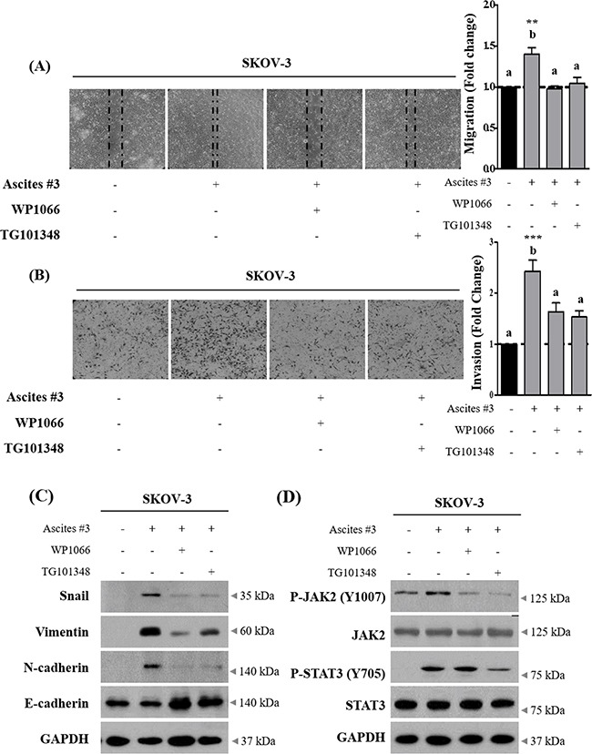
Figure 4. Inhibition of JAK2-STAT3 signaling suppress ascites-induced migration and invasion in SKOV-3 cells.

A. SKOV-3 cancer cells were treated with 10% ascites, with or without JAK2 and STAT3 inhibitors, WP1066 and TG101348. After 24 hr, wound healing ability was verified by measuring wound closed area under a light microscope (magnification x40). B. SKOV-3 cancer cells were seeded into the upper chamber of Matrigel-coated membrane in transwells. Cell invasion were induced by ascites with or without JAK2 and STAT3 inhibitors. After 24 hr, invaded cells at the bottom of the transwell were stained with 0.5% crystal violet and counted under a light microscope (magnification x200). C. SKOV-3 cancer cells were treated with JAK2 and STAT3 inhibitors as above. After 24 hr, the expression of Snail, Vimentin, N-cadherin and E-cadherin were examined by western blot. GAPDH was used as an internal control. D. SKOV-3 cancer cells were treated as above. The expression of p-JAK2 (Y1007), JAK2, p-STAT3 (Y705) and STAT3 were examined by western blot. GAPDH was used as an internal control. ** and *** represent P < 0.01 and P < 0.001, respectively.
