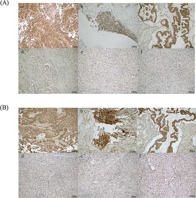
Figure 2. Immunohistochemical staining of CLIC1 and LGALS3BP in ovarian cancer and normal ovary tissues.

A. CLIC1 was positive staining in ovarian cancer cells (a, b and c), as well as benign ovarian epithelial cells were with negative or weak CLIC1 staining (d, e and f); B. LGALS3BP was strong positive staining in ovarian cancer cells (a, b and c) and almost negative in normal ovarian epithelial cells (d, e and f). Original magnification: 200×.
