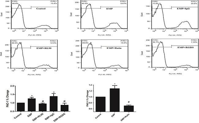
Figure 7. PDI was involved in EMP-induced platelet GPIIb/IIIa activity.

Incubate EMPs with 1μg/ml RL90, IgG (1μg/mL), rutin(10μM), rutin(60μM) at 37°C for 30min respectively, then stimulate platelet with the EMPs. Or incubate EMPs with RGDS (10μg/mL) with platelet at 37°C for 15min, then incubate the platelet with EMPs. Detect platelet expression of PAC-1 (represented activated GPIIb/IIIa) by flow cytometry. Histograms represent the PAC-1% change. *P < 0.05 compared with control, #P < 0.05 compared with EMPs treatment. Data are means ± SD from at least three separate experiments.
