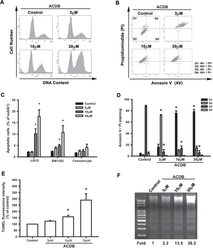
Figure 2. ACDB induces apoptosis in human chondrosarcoma cells.

A and C. Cells were treated with vehicle or ACDB for 48 h. The cell cycle analysis (PI staining) was examined and quantified by flow cytometry. B and D. JJ012 cells were treated with vehicle or ACDB for 48 h. The percentage of apoptotic cells was analyzed by flow cytometry of Annexin V/PI double staining, and the results were quantified. E. Cells were treated with vehicle or ACDB for 48 h. The TUNEL positive cells were examined by flow cytometry (n = 4). F. For measuring apoptosis, the DNA fragmentation assay was performed as described in Materials and Methods section. Results are expressed as the means ± SEM of four independent experiments. * P < 0.05 as compared with control group.
