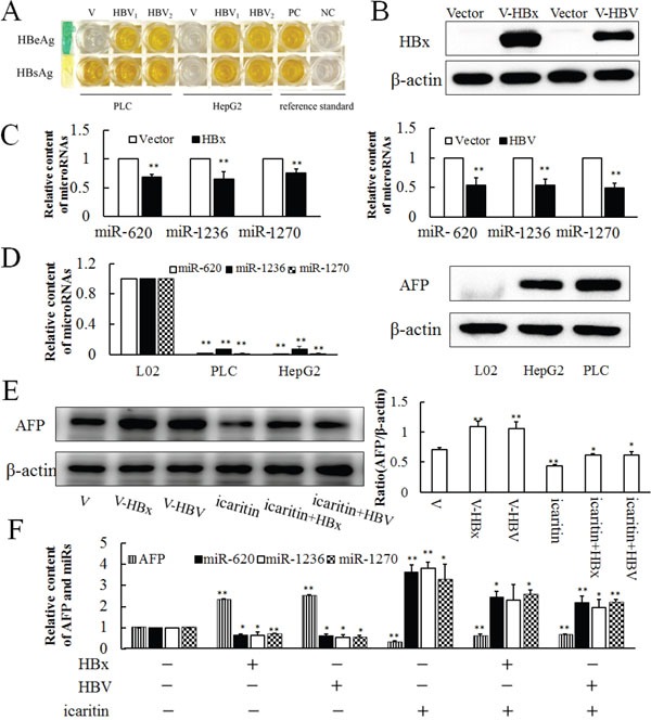
Figure 5. Expressions of miR-620, miR-1236, miR-1270 in HBV- and HBx-transfected cells.

A. HBsAg and HBeAg were detected in the cell culture medium by ELISA after 36 h of infection with HBV in PLC and HepG2 cells. PC. Positive control. NC. Negative control. B. Western blotting analysis of HBx and HBV transfected cell lines. C. Effects of HBV and HBx transfection on miR-620, miR-1236, miR-1270 expressions after 36 h. D. Background expressions of miR-620, miR-1236, miR-1270 and AFP in L02, PLC and HepG2 cell lines analyzed by qRT-PCR and western blotting. E. Effect of HBV and HBx infection cooperate with icaritin on AFP expression analysis by western blotting (E) and qRT-PCR F. The panel on the right side of the images is the densitometric analysis. The each image is representative of at least three independent experiments. Data represents mean ± SD of three samples. *P < 0.05 and **P < 0.01 as compared with controls.
