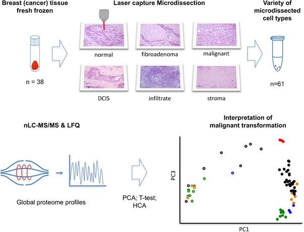
Figure 1.

Schematic representation of the LCM‐proteomics workflow. Fresh frozen breast tissues were subjected to LCM, from which both epithelial and stromal cell regions were collected. Proteins were extracted, trypsin digested, and subjected to nano‐LC‐MS/MS analysis, after which the protein abundance data were analyzed by PCA.
