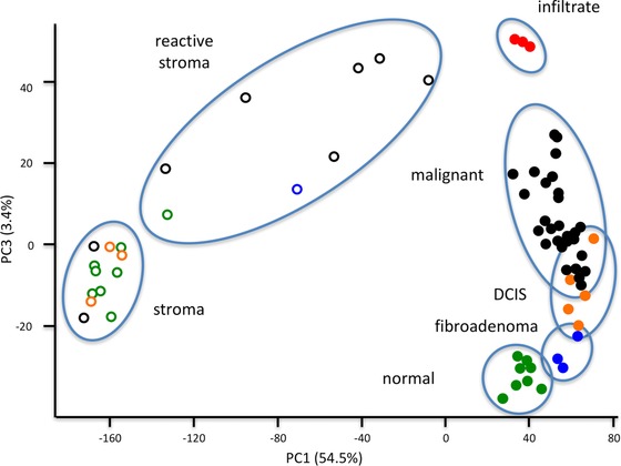
Figure 3.

PCA scores plot of principal components 1 and 3. Microdissected samples clustered according to their histology, with principal component 1 discriminating between stroma and epithelium and principal component 3 discriminating between malignancy, on the basis of expression of immune regulatory proteins. Red filled squares: microdissected infiltrate from malignant tumor sections; black filled circles: microdissected malignant tumor epithelium; orange filled circles: microdissected ductal carcinoma in situ epithelium; blue filled circles: microdissected epithelial cells from fibroadenoma sections; green filled circles: microdissected epithelial cells from histologically normal sections. Black circles: stroma dissected from histologically malignant sections; blue circles: stroma dissected from a fibroadenoma section; orange circles: stroma dissected next to ductal carcinoma in situ lesions; green circles: stroma dissected from histologically normal sections.
