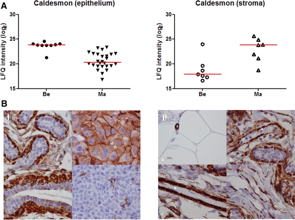
Figure 5.

Protein abundance and immunostaining of caldesmon. (A) Protein abundance in benign and malignant epithelium and stroma. (B.I) Cytoplasm and membrane staining in; (a) myoepithelial layer of normal acini; (b) myoepithelial layer of a normal duct (some apical staining in luminal layer); (c) positive invasive breast tumor cells; (d) endothelial cells of a negative invasive breast tumor. (B.II, a) positive staining in capillaries, negative in fat cells; (b) myoepithelial layer in normal glands (negative in inflammatory cells (⇑) and tumor cells (↑)); (c) pericytes (negative in endothelial cells in the bloodvessel (▲); (d) positive staining in fibroblasts.
