
Figure 1.
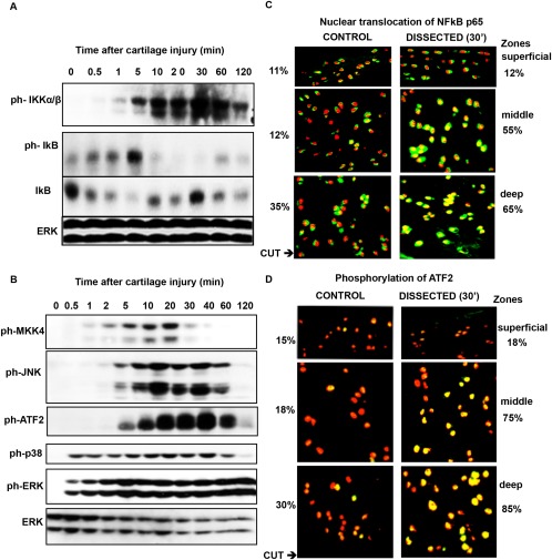
Activation of the NF‐κB and JNK pathways and their upstream regulators in injured porcine cartilage. Porcine metacarpophalangeal joints were equilibrated at 37°C for 1 hour. Articular cartilage was dissected and either snap‐frozen or kept in serum‐free medium for the indicated times. A and B, Cartilage was washed with 1× phosphate buffered saline and lysed in radioimmunoprecipitation assay buffer. Lysates were analyzed by Western blotting for phospho‐IKK (ph‐IKK), phospho‐IκB, and IκB (A) and for phospho‐MKK‐4, phospho‐JNK, phospho–activating transcription factor (phsopho–ATF‐2), phospho‐p38, and phospho‐ERK (B). ERK was blotted to check that the lanes were equally loaded. Results are representative of several replicate experiments. C and D, After incubation, cartilage was embedded in OCT, and cryosections were stained with antibodies against the p65 subunit of NF‐κB (C) or phospho‐ATF‐2 (D). The localization of the 2 proteins in different cartilage zones was determined using confocal microscopy. In each cryosection, a total of 500 cells were counted (100 cells in the superficial and deep zones and 300 in the middle zone). Percentages are the proportion of activated cells in the different zones. Representative images are shown. Original magnification × 60.