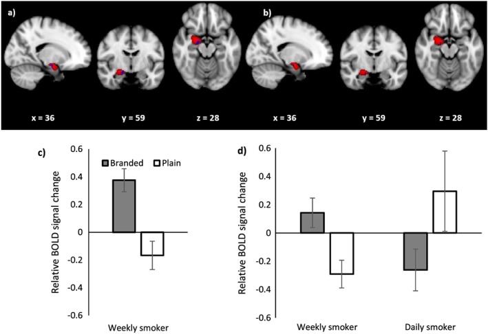
Figure 4.

(a) Blood oxygen level‐dependent (BOLD) activation in the right amygdala for the comparison plain > branded controlling for fixations to health warnings (i.e. eye‐tracking weighted analysis) among weekly smokers and (b) weekly smokers compared with daily smokers. Coordinates represent coordinates in Montreal Neurological Institute‐152 (MNI‐152) coordinate space. The red region shows the extent of the 30% threshold applied to the probabilistic right amygdala mask and the blue region is the activated cluster. (c) Relative BOLD signal change in the activated region of the right amygdala for the equally weighted analysis for the comparison branded > plain for weekly smokers. (d) Relative BOLD signal change in the activated region of the right amygdala for the equally weighted analysis for the comparison weekly smokers > daily smokers. Error bars represent the standard error of the mean
