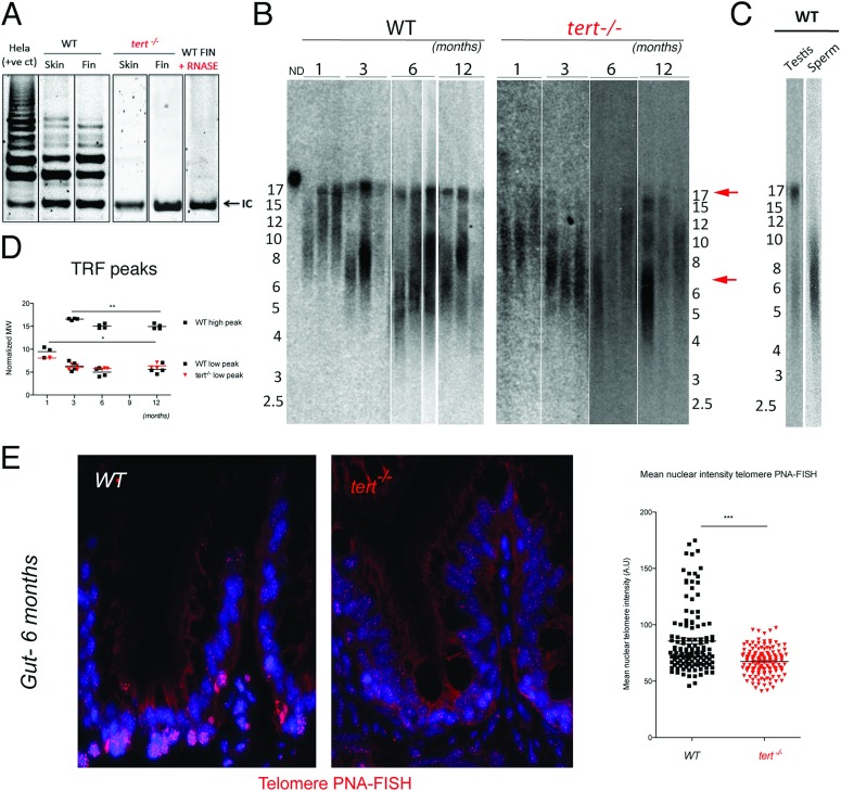
Fig 1. Telomerase mutant zebrafish have shorter telomeres than WT siblings.
A) Representative image of TRAP assay showing that telomerase is not active in the tert−/− zebrafish, as compared to tert+/+ siblings. Here shown are caudal fin and skin protein extracts. Hela cell extract is shown as positive control. N = 4. B) Representative image of restriction fragment analysis of caudal fin genomic DNA of 3 different individuals at different ages, by southern blot (random primer-labelled telomeric probe (CCCTAA)12 32P-dCTP). tert+/+ Zebrafish have heterogeneous telomeres, with two distinct peaks of different lengths. In tert+/+ the highest peak (∼16 Kb, top red arrow) becomes more distinct after 1 months of age and decreases in length over-time (B and D). The lowest peak of telomere intensity also decreases in length (bottom red arrow, B and D). tert−/− zebrafish have shorter telomeres than tert+/+ siblings in different tissues (see also Figure S1A and S1B), observed by the decrease in length of the higher TRF peak. The shortest TRF peaks accompany those of tert+/+ siblings, and decrease over-time at similar rates. C) Testes fractionation in tert+/+ reveals the two-telomere length populations in whole testes, whereas mature sperm only shows the shorter TRF smear of about 6 Kb, suggesting different telomere lengths in different cells within a tissue. D) TRF mean sizes were calculated as described in [50]. E) Telomere PNA-FISH in 6-month-old gut tissue shows cells with different telomere intensities in the wild type, mainly localizing to the proliferative niche. In contrast tert−/− mutants display cells with less bright and more homogeneous telomere intensity.

