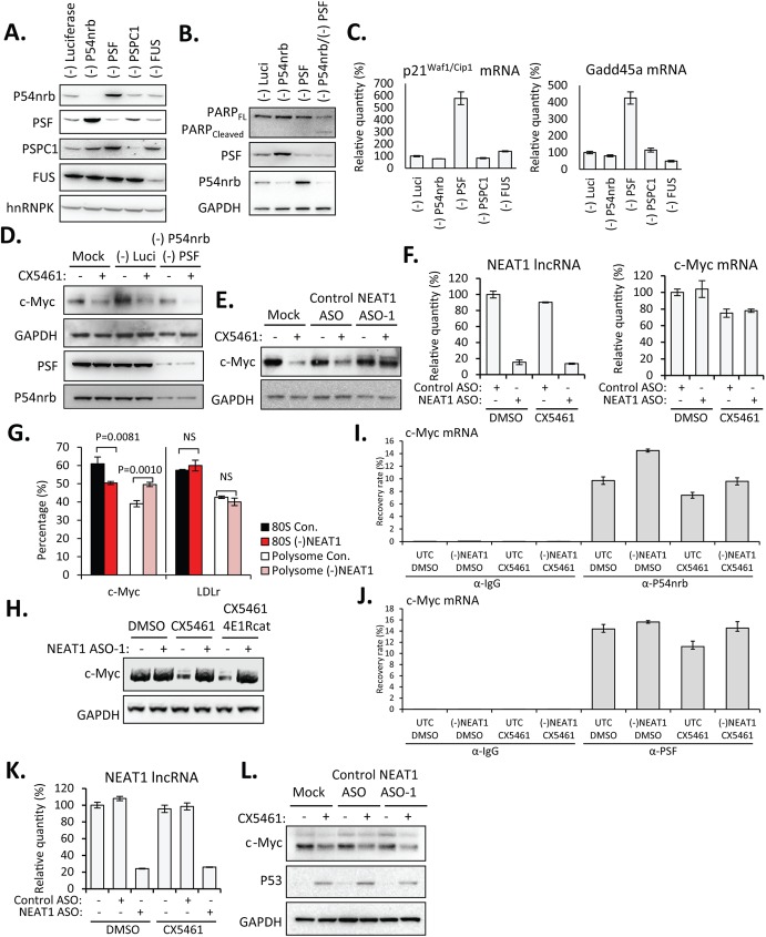
Fig 5. Depletion of NEAT1 lncRNA allowed increased association between P54nrb/ PSF and c-Myc mRNA to facilitate the IRES-translation of c-Myc.
A. Western analysis of protein reduction by corresponding siRNAs. B. Simultaneous depletion of P54nrb and PSF by siRNAs caused proteolytic cleavage of PARP. C. qRT-PCR suggested the upregulation of p21Waf1/Cip1 and Gadd45a mRNAs upon depletion of PSF. D. Simultaneous depletion of P54nrb and PSF by siRNAs reduced levels of c-Myc protein in the presence of CX5461. HeLa cells were transfected with individual siRNAs at a final concentration of 3 nM for 48 hrs, followed by the treatment with DMSO or 125 nM CX5461 for 6 hrs. E. Depletion of NEAT1 lncRNA rescued levels of c-Myc protein in the presence of CX5461 in HeLa cells. HeLa cells were mock-transfected or transfected with 30 nM NEAT1 or control ASOs for 2hrs, followed by the treatment with DMSO or 125 nM CX5461 for 6 hrs. F. qRT-PCR showed levels of NEAT1 lncRNA and c-Myc mRNA in HeLa cells. G. qRT-PCR for relative levels of c-Myc and control LDLr mRNAs in pooled polysome (F12-F25) and mono-ribosome (80S) (F6-F10) fractions from CX5461-treated HeLa cells transfected with NEAT1 ASO-1 or control ASO (con.), as in S5 Fig). Error bars indicate s.d. of three independent experiments. P values were calculated based on unpaired t-test. NS, not significant. H. HeLa cells were mock-transfected or transfected with 30 nM NEAT1 for 2 hrs, followed by the treatment with DMSO, CX5461 (125 nM), or CX5461 (125 nM)/4E1Rcat (10 μM) for 6 hrs. I and J. qRT-PCR of levels of c-Myc RNA co-immunoprecipitated with anti-P54nrb (I.) or anti-PSF (J.) antibodies. Immunoprecipitation by anti-mouse IgG antibody serves as a background control. Results are presented as percent recovery from the input material. K. qRT-PCR showed levels of NEAT1 lncRNA in MCF7 cells. L. Depletion of NEAT1 lncRNA failed to rescue the levels of c-Myc protein in the presence of CX5461 in MCF7 cells. Experiments were performed as described in Fig 5E.

