Figure 3.

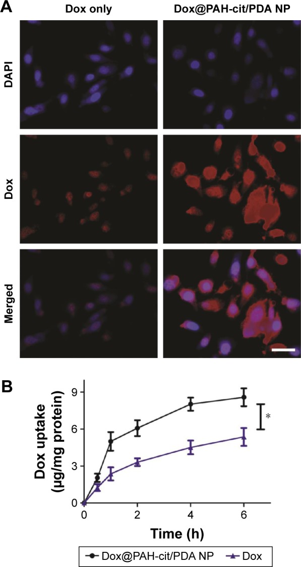
(A) Cellular uptake of free Dox and Dox@PAH-cit/PDA NPs in PC3 cells revealed by CLSM images; scale bar =50 µm. (B) Quantitative analysis of intracellular concentration of Dox after cells incubated with free Dox or Dox@PAH-cit/PDA NPs. The ratio between Dox and cell protein concentration was detected. *P<0.05 was calculated by using Student’s t-test.
Abbreviations: CLSM, confocal laser scanning microscopy; DAPI, 2-(4-amidinophenyl)-6-indolecarbamidine dihydrochloride; Dox, doxorubicin; NP, nanoparticle; PAH-cit, poly(allylamine)-citraconic anhydride; PDA, polydopamine.
