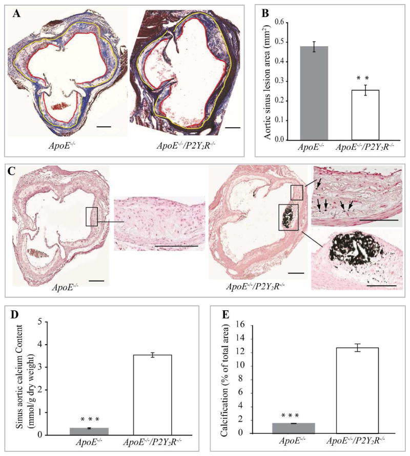
Fig. 1. P2Y2 receptor deficiency reduces atherosclerosis and increases calcification of atherosclerotic lesions.
(A) Light micrographs of representative Masson’s trichrome stained aortic root lesions from ApoE−/− and ApoE−/−/P2Y2R−/− mice, showing atherosclerotic lesion (area between yellow and red lines) as well as collagen and muscle fibers in the lesions. (B) Quantitative analysis of lesions in the aortic sinus. Data represent the mean ± SEM lesion area for 5 consecutive sections in each of the 12 mice examined for each genotype. (*p<0.001). Scale represents 100 μm. Arrows indicate lesions. (C) Representative images of sections stained with Von Kossa to detect calcification. Areas in insert have been magnified. Arrows indicate spotty calcium deposits. Scale bar = 100μm. (D) Quantification of the sinus aortic calcium content expressed as mmol/g of dried weight and (E) calcification presented as percentage of Von Kossa-positively stained areas in the total aortic lesion. Quantification was done using 5 cross sections in each of the 12 ApoE−/− mice and 12 ApoE−/−/P2Y2R−/− mice. Bar values are means ± SEM, *p<0.001.

