
Figure 3.
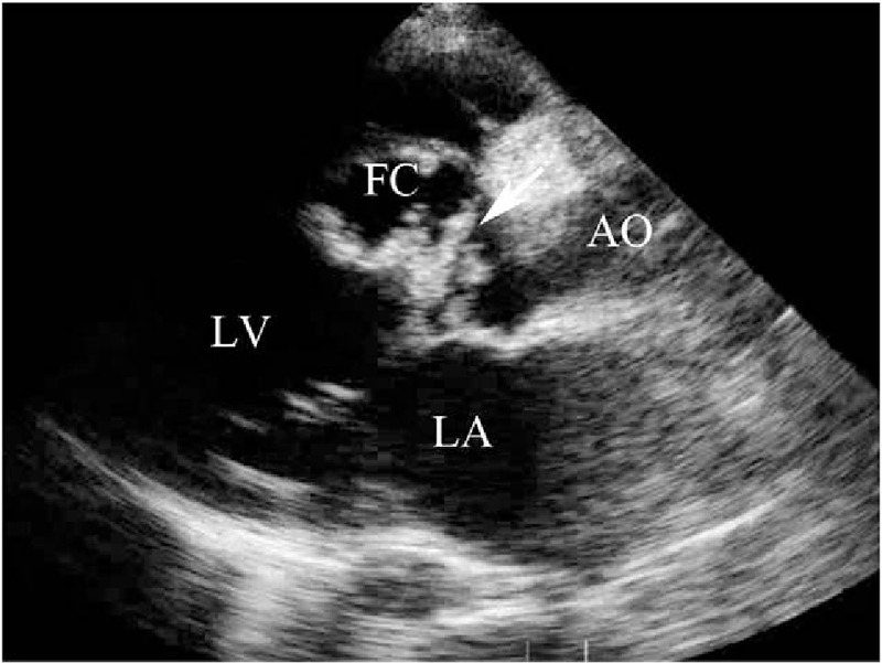
(Patient #7) Transthoracic echocardiogram (parasternal long axis view) showing thickened aortic valve leaflets with vegetations and the entry site of rupture (arrow) from the right sinus of Valsalva into the dissecting cystic-like false cavity in the interventricular septum (see also video 1). AO = aorta, FC = false cavity, LA = left atrium, LV = left ventricle.