
Figure 4.
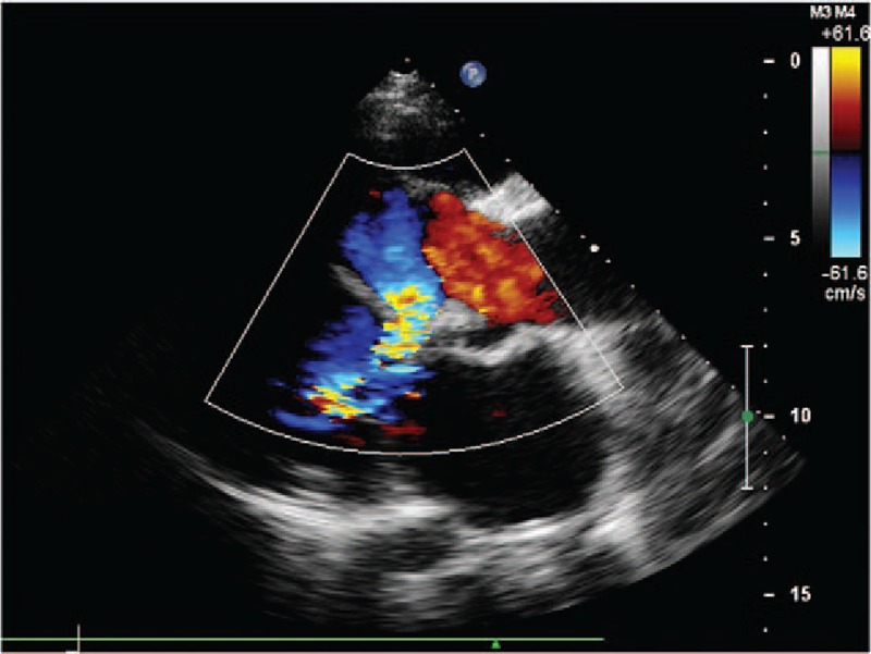
(Patient #7) Transthoracic echocardiogram (parasternal long axis view with color) showing both the communication between the right sinus of Valsalva aneurysm and the dissecting cavity in the interventricular septum and the communication between the dissecting cavity in the interventricular septum and the left ventricle (see also video 2).