
Figure 5.
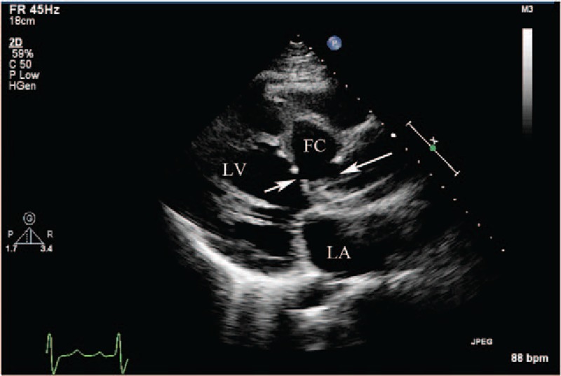
(Patient #11) Transthoracic echocardiogram (parasternal long axis view) showing a mechanical aortic valve in situ with annular detachment and the entry site of rupture (arrow on right) from the right sinus of Valsalva aneurysm into the dissecting false interventricular septal cavity, and the entry site of rupture (arrow on left) from the false interventricular septal dissecting cavity into the left ventricular cavity. (See also video 3). FC = false cavity, LA = left atrium, LV = left ventricle.