
Figure 6.
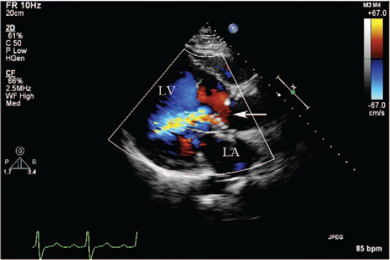
(Patient #11) Transthoracic echocardiogram (parasternal long axis view with color) showing the communication (arrow) from the right sinus of Valsalva aneurysm into the interventricular septal cavity, and the entry site of rupture from the false interventricular septal dissecting cavity into the left ventricular cavity. LA = left atrium, LV = left ventricle.