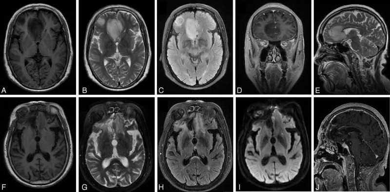
Figure 3.

Brain magnetic resonance imaging showed a partial response of glioblastoma in a 46-year-old male patient before surgery and after 7 cycles of chemotherapy. (A) Preoperative, axial view, T1-weighed image (T1WI). (B) Preoperative, axial view, T2-weighed image (T2WI). (C) Preoperative, axial view, fluid attenuation inversion recovery image (FLAIR). (D) Preoperative, coronal view, contrast-enhanced T1WI. (E) Preoperative, sagittal view, T2WI. (F) Postoperative, axial view, T1WI. (G) Postoperative, axial view, T2WI. (H) Postoperative, axial view, FLAIR. (I) Postoperative, axial view, diffusion-weighted imaging. (J) Postoperative, sagittal view, contrast-enhanced T1WI.
