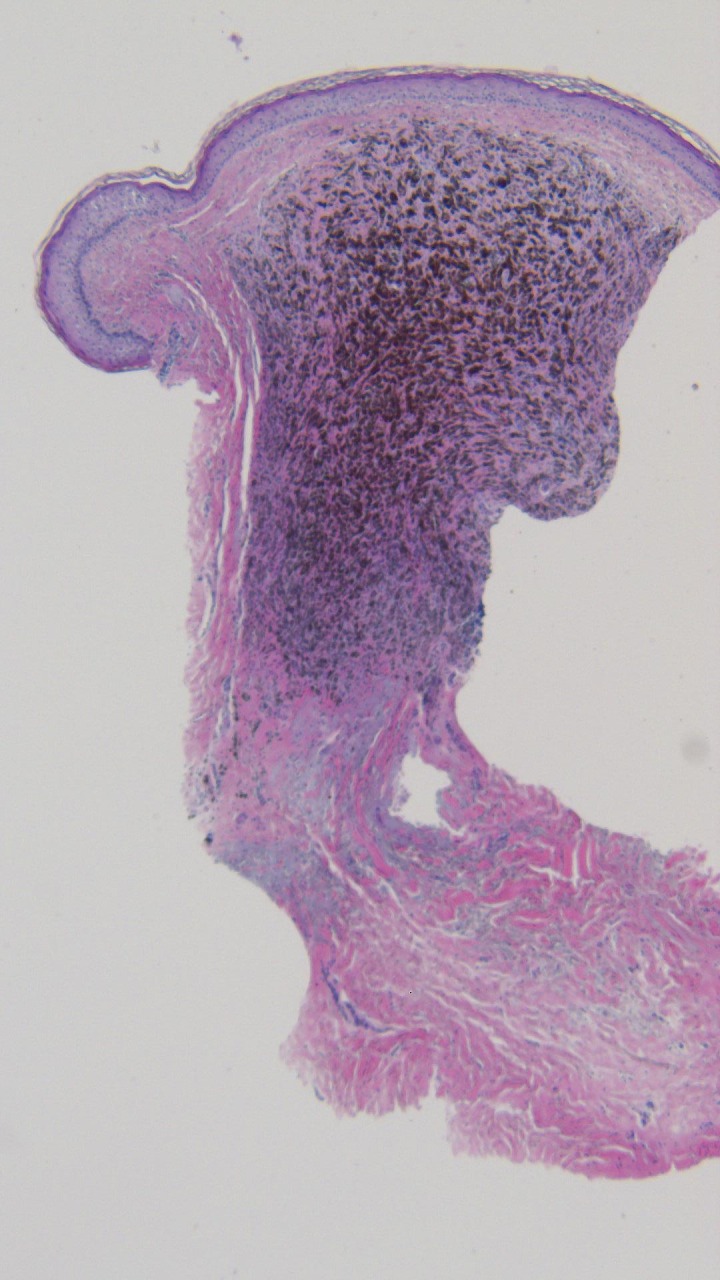
Figure 13. Microscopic examination of the punch biopsy of the tumoral melanosis on the left parietal scalp.

Dense collections of melanophages are seen in a nodular aggregate; tumor melanocytes are not observed (hematoxylin and eosin; x = 4).

Dense collections of melanophages are seen in a nodular aggregate; tumor melanocytes are not observed (hematoxylin and eosin; x = 4).