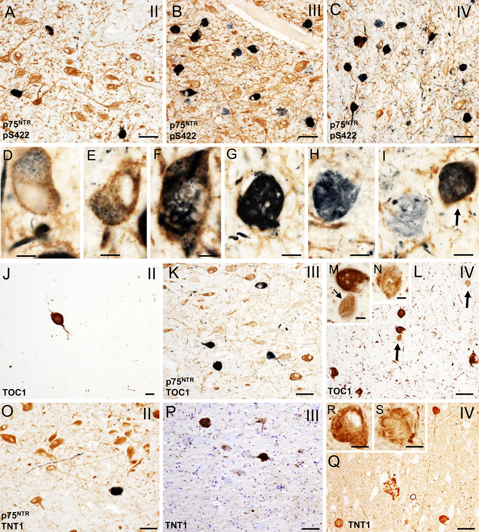
Figure 2.
A–C. Photomicrographs of tissue dual immunostained for p75NTR (brown) and pS422 (dark blue/black) in the anterior subfields of the nbM showing the presence p75NTR containing pS422 inclusions (dark blue) and neuropil threads (dark blue) in CTE stages II (A), III (B) and IV (C). Note that many more double p75NTR/pS422 positive neurons and neuropil threads (dark blue) were seen in stage III and IV compared to stage II. D–G. High power images of double immunolabeled p75NTR/pS422 neurons in the anteromedial nbM showing intraneuronal cytoplasmic pS422 imunoreactivity (black) containing granular (D and E), filamentous (F) and skein of yarn-like (G) phenotypes in a stage IV CTE case. H and I. Twisted filamentous p75NTR positive inclusion (blue) resembling a skein of yarn in the nbM neuropil. Arrows indicates a double immunolabeled p75NTR/pS422 neuron in I. J–L. Photomicrographs of single TOC1 (brown) (J and L) and dual immunolabeled p75NTR/TOC1 (dark blue/black) neurons and neuropil threads in anterior subfields of the nbM in a stage II (J), III (K) and IV (L) CTE cases. Note the increase in the extent of TOC positive neuropil threads in stage IV. M and N. Insets showing higher magnification images of granular (small arrow), compact spherical (M) and twisted-filamentous (N) TOC1 immunoreactive profiles (brown) in the anterormedial subfield of the nbM neurons from panel L (arrows). O–Q. Photomicrographs of double immunolabeled TNT1/p75NTR (dark blue) (O) and single reactive TNT1 (brown) (P and Q) positive neurons and neuropil threads in the anterior subfields of the nbM from a stage II (O), III (P) and IV (Q) CTE cases. R and S. Insets showing details of the granular and filamentous TOC1 immunoreactivity in anteromedial subfield of the nbM neurons from a stage IV CTE case. Tissue was counterstained with cresyl violet in panel P. Scale bars: 75 µm (A–C), 65 µm (L, O, Q), 50 µm (K, P), 25 µm (J), 15 µm (M, R, S), 12 µm (G), 10 µm (D, E, H) and 8 µm (F, I, N).

