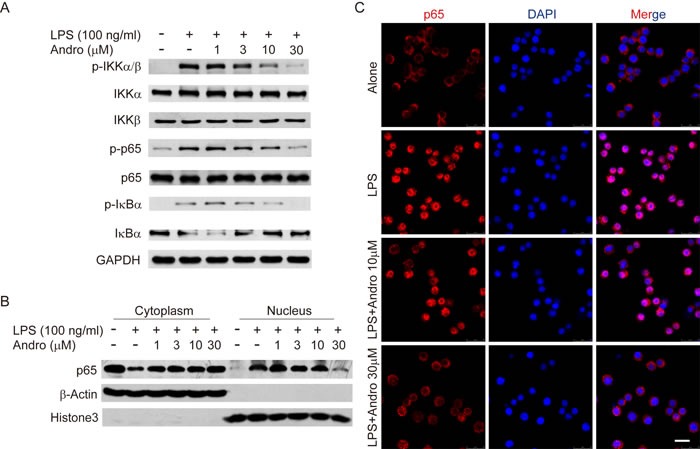
Figure 7. Andrographolide inhibits LPS-induced activation of NF-κB in RAW264.7 cells.

Cells were treated with various concentrations of Andrographolide in the absence or presence of LPS for 20 mins. A. The protein levels of phosphorylated and total IKKα, IKKβ, p65 and IκBα were determined by western blot. B. p65 levels in the cytosol and nucleus were assayed. β-actin and Histone3 were shown as loading controls. C. The subcellular localization of p65 was assayed by Immunofluorescence via confocal microscope. Nuclei were stained by DAPI. In all experiments, the representative data are shown of at least 3 independent experiments. Scale bar 20 μm.
