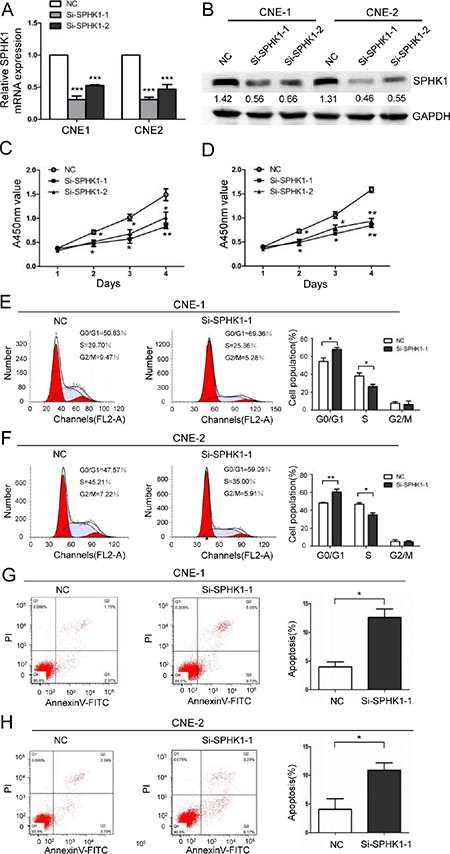
Figure 1. The downregulation of SPHK1 inhibits NPC cell proliferation and induces cell cycle arrest and apoptosis.

(A) CNE-1 and CNE-2 cells were transiently transfected with SPHK1 siRNAs using Lipofectamine. SPHK1 mRNA expression was analyzed using qRT-PCR at 24 h after transfection. β-actin was used as an internal control. (B) Western blot analysis showing the protein level of SPHK1 in CNE-1 and CNE-2 cells that were transfected with si-SPHK1-1 or si-SPHK1-2. After 48 h, SPHK1 levels were lower in the treated cells than in the negative control (NC). GAPDH was used as a loading control. (C, D) The proliferation of CNE-1 and CNE-2 cells was measured at the indicated times after transfection using a Cell Counting Kit-8 (CCK-8) kit. The results are presented as the means ± S.D. of values that were obtained in three independent experiments. Significance was calculated using Student's t-tests. (E, F) Flow-cytometric analysis of CNE-1 and CNE-2 cells that were infected with NC and SPHK1 siRNAs. (G, H) CNE-1 and CNE-2 cells were transfected with NC or SPHK1 siRNAs, stained with propidium iodide and Annexin V-fluorescein isothiocyanate (FITC), and analyzed using flow cytometry. Silencing SPHK1 significantly increased the rate of apoptosis in both cell types. *P < 0.05, **P < 0.01.
