
Figure 12. Klotho expression in human melanoma tissues and its clinical significance.
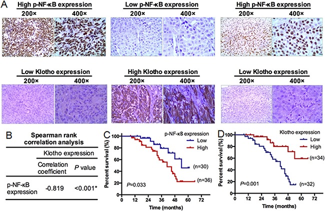
Sixty-six melanoma tumor tissues were subjected to immunohistochemical staining of p-NF-κB and Klotho protein. A. Representative immunohistochemical staining of p-NF-κB and Klotho expression. Left panels: high p-NF-κB expression, low Klotho expression (200x, 400x) in melanoma tissues. Middle panels: low p-NF-κB expression, high Klotho expression (200x, 400x) in melanoma tissues. Right panels: high p-NF-κB expression and low Klotho expression (200x, 400x) in melanoma tissues with ulceration. B. The spearman rank correlation analysis between the expression of Klotho and p-NF-κB. C. Kaplan-Meier analysis of survival in patients with high or low p-NF-κB levels. N=66. D. Kaplan-Meier analysis of survival in patients with high or low Klotho expression. N=66.