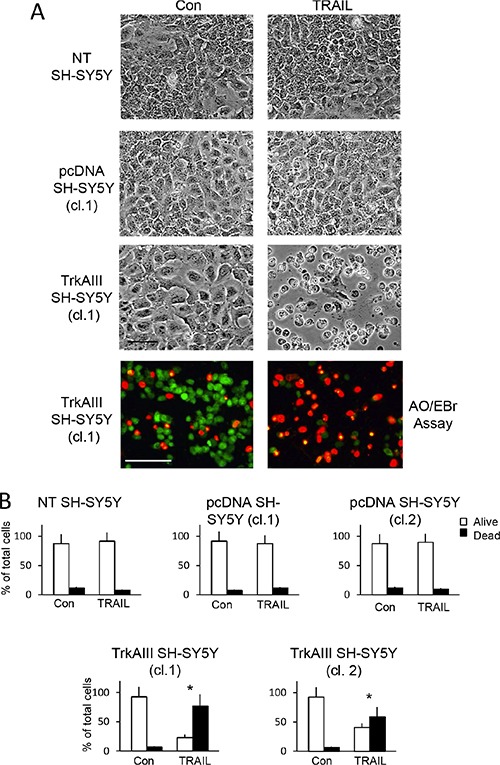
Figure 1. TRAIL induces apoptosis of TrkAIII SH-SY5Y cells.

(A) Representative phase contrast (black and white) and fluorescent (green and orange) micrographs (Bar = 100 μm) demonstrating marked induction of TrkAIII SH-SY5Y but not non-transfected (NT) SH-SY5Y or pcDNA SH-SY5Y cell death, following 24 hours incubation with TRAIL (200 ng/ml). (B) Histograms displaying the mean (± SD) percentage (%) survival (white) and death (black) cells of NT SH-SY5Y, independent pcDNA SH-SY5Y clones (cl.1 and 2) and independent TrkAIII SH-SY5Y clones (cl.1 and cl.2) incubated for 24 hours with TRAIL (200 ng/ml), in three independent cell death assays each performed in duplicate (* = statistical significance with respect to Con).
