
Figure 6. Combined use of ER stress inducer and autophagy late stage inhibitor to restore the ICD inducer activity of 5-Fu and SN-38.
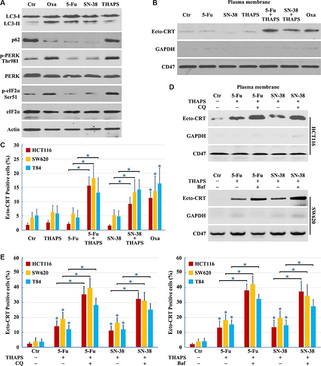
(A) HCT116 cells treated with 10 μg/ml Oxaliplatin (Oxa), 10 μg/ml 5-Fu, 80 nM SN-38 or 500 nM THAPS for 8 h were lysed and subjected to immunoblot detection; (B–C) Cells were treated with 10 μg/ml Oxa, 10 μg/ml 5-Fu, 80 nM SN-38, 500 nM THAPS, 10 μg/ml 5-Fu plus 500 nM THAPS or 80 nM SN-38 plus 500 nM THAPS for 8 h followed by Ecto-CRT detection (B) or send to immunofluorescence detection and quantification of Ecto-CRT (C); (D–E) Cells were treated with 10 μg/ml 5-Fu plus 500 nM THAPS, 80 nM SN-38 plus 500 nM THAPS, 10 μg/ml 5-Fu plus 500 nM THAPS plus 50 μM CQ/ 50 nM Bafilomycin A1 (Baf) or 80 nM SN-38 plus 500 nM THAPS plus 50 μM CQ/ 50 nM Baf for 8 h followed by Ecto-CRT detection (D) or send to immunofluorescence detection of Ecto-CRT (E). Results are representative of three independent experiments. The values represent the mean ± S.E. of at least three independent experiments. * denotes p < 0.05.