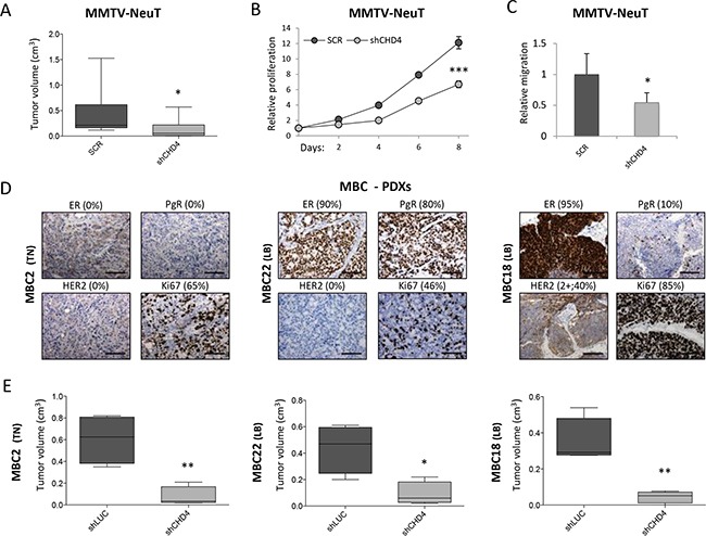
Figure 3. CHD4 role in the MMTV-NeuT transgenic mouse and in PDX breast cancer models.

A. Cells derived from dissociation of spontaneous mammary tumors of MMTV-NeuT mice were infected with two pooled shRNAs silencing Chd4 gene and a corresponding control (scramble – SCR) and subsequently transplanted in FVB mice. Box plots represent the tumor volume (mean±SD - cm3) of eight to ten distinct tumors grown in vivo. Statistical difference between groups was calculated using Mann-Whitney U test (U=14.0; *: P<0.05). Transduced cells were also used to analyze in vitro cell proliferation (eight days) B. and migration C. Statistical significance was calculated by applying a Student t-test (*: P<0.05; ***: P<0.001). D. Immunohistochemical staining of Estrogen (ER), Progesterone (PgR), HER2+ receptors and Ki67 in three metastatic breast cancer (MBC) patient-derived xenografts (PDXs). Percentage (%) of positive cells is reported for each staining. Scale bar: 100μm. E. Box plots representing tumor volume (mean±SD - cm3) of four distinct tumors arisen after transplantation of PDXs cells infected with a pool of two shRNAs targeting CHD4 and the corresponding control (shLUC) in NSG mice. Statistical significance was calculated by applying a Student t-test (*: P<0.05; **: P<0.01).
