
Figure 4. Efficient infection of cells, with the GFP producing GLV-1h68, leads to apoptotic cell death due to the loss of the anti-apoptotic MCL-1 protein and the inhibitors of apoptosis (IAPs).
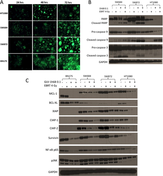
A. GFP transgene expression in the rat BN175 cell line and a panel of human sarcoma cell lines (HT1080, SW684 and SW872) at 24, 48 and 72 hours after treatment with GLV-1h68 (MOI 1). B. Western blot analysis of 3 human sarcoma cell lines after treatment with GLV-1h68 (MOI 0.1) and EBRT (4 Gy). C. Western blot analysis of the expression of anti-apoptotic BCL2 proteins, IAPs and downstream pro-survival signalling pathways 48 hours after treatment with GLV-1h68 (MOI 0.1) and EBRT (4 Gy) alone or as combination therapy. The MCL-1 protein was consistently detected at 40kDa in the human cell lines and at 37kDa in the BN175 rat cell line.