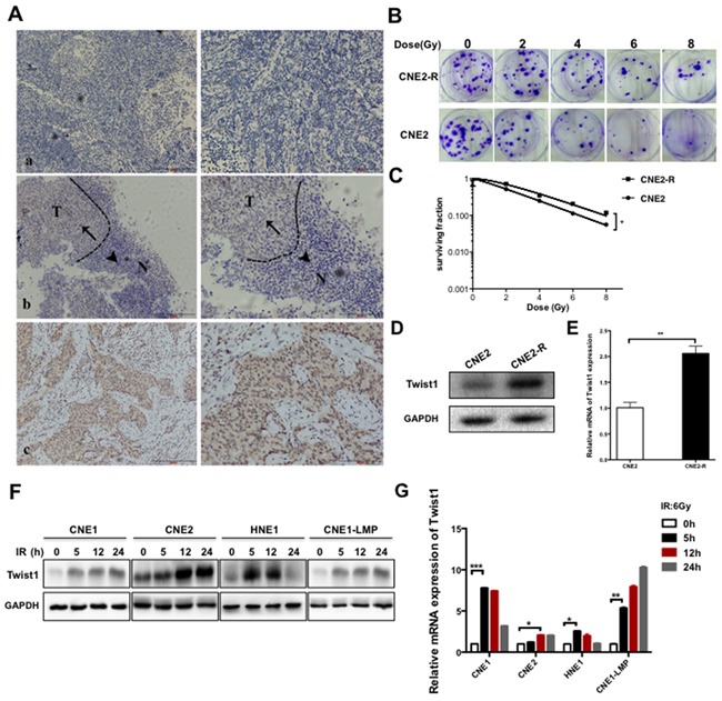
Figure 1. Twist1 is correlated with malignance and radioresistance of NPC.

A. The representative immunohistochemical (IHC) stainings for Twist1 in paraffin-embedded tissue sections. a, negative stainings of Twist1 in nasopharyngeal inflammation tissues. b, the different expressions of Twist1 in normal (N) and tumorous (T) tissues, as indicated by arrows. c, positive stainings of Twist1 in nasopharyngeal cancer. Photos were taken under 10X, 200X magnification respectively. B. The representative colony formation images of radiation resistant CNE2 cells (CNE2-R) and CNE2 cells, survival curves were prepared in C. the error bars represent means ± standard deviations (SDs) from 3 independent experiments, *P<0.05 versus (vs.) control. D. The expressions of Twist1 were showed by immunoblot (IB) in CNE2 and CNE2-R. E. The quantification of Twist1 mRNA in CNE2 and CNE2-R cells, the error bars are means ± SDs from 3 independent experiments, **P<0.01 vs. control. F. The IB analysis of WCLs (whole cell lysates) derived from different NPC cell lines after exposure to radiation specific for Twist1. G. The quantification and analysis of Twist1 mRNA in different NPC cell lines irradiated with 6 Gy radiation, the error bars are means ± SDs from 3 independent experiments, *P<0.05 vs. control;**P<0.01 vs. control; ***P<0.005 vs. control.
