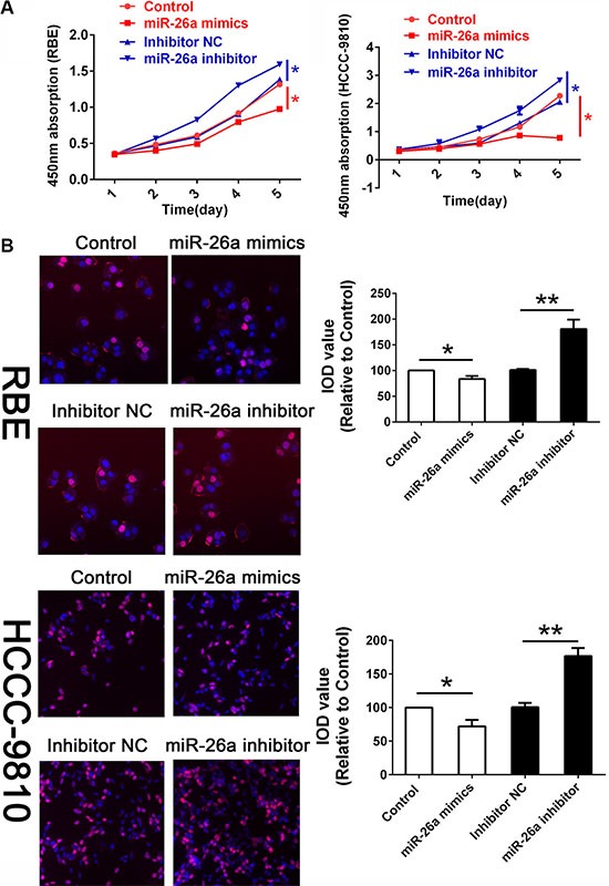
Figure 2. miR-26a suppressed cell proliferation in vitro.

(A) CCK8 assay were employed to detecting the proliferation of CCA cells. Data was presented with Mean ± SE. *indicates significant difference compared with that of control cells (P < 0.05). (B) The EDU stain also performed in cells treated as described above with a magnification of 200. The integral optical density value of cells treated with control plasmids was normalized to 100%.
