
Figure 1. Establishment of NCI-H1975 cells resistant to OSI.
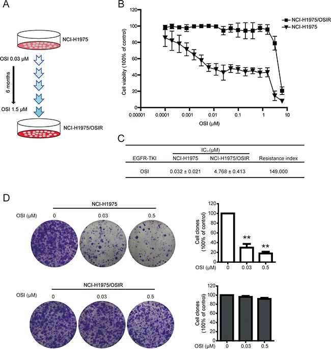
A. Schematic of establishing OSI-resistant NCI-H1975 cells. B. Cells were incubated with various concentrations of OSI for 72 h. The anti-proliferative effects of OSI in NCI-H1975 and NCI-H1975/OSIR cells were evaluated by MTT assay. *P<0.05 and **P<0.01, compared with the 0 μM OSI treatment. C. The IC50 values of OSI in NCI-H1975 and NCI-H1975/OSIR cells. D. Cells were exposed to OSI for 72 h and incubated with drug-free medium for 7 days. Then, cells were fixed with 4% PFA and stained with crystal violet. The cell colonies were photographed and representative images were exhibited. For quantitative assay, clones were dissolved in acetate acid after crystal violet staining and absorbance was recorded. *P<0.05 and **P<0.01.