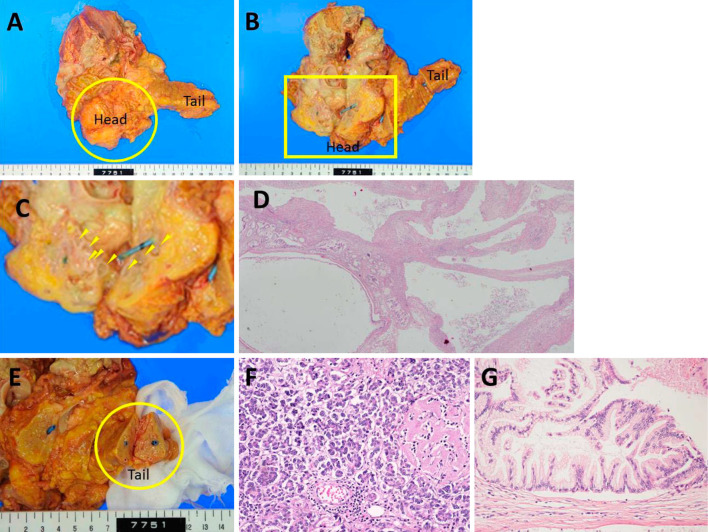
Figure 6.
The pathological findings. A: An elastic soft mass (6×8×5 cm, circle) extended from the pancreatic head and surrounded Gerota’s fascia of the right kidney. B: The cut surface of the pancreatic head showed the presence of cystic lesions of various sizes, with the leakage of milky-white viscous fluid (arrowhead). The rupture point of the IPMN could not be located due to the presence of inflammation, necrosis, and fibrosis. A pancreatic stent had been placed in the main pancreatic duct. C: An enlarged view of the square area of B. D (×20): The histological findings of pancreatic head showed the presence of cystic lesions of various sizes. E: Macroscopic findings showed that the structures of the tail (circle) of the pancreas were relatively well preserved. F (×200): The histological findings showed that the acinar structures of the tail of the pancreas were relatively well preserved; small regions of fat necrosis were sporadically found and hyalinization of the islets of Langerhans had occurred due to diabetes mellitus. No fibrosis was found in the tail of the pancreas. The macroscopic and histological findings showed that there was no obvious chronic pancreatitis. G (×200): Atypical papillary proliferation was found in some of the cystic lesions at the pancreatic head, indicating the presence of an intraductal papillary mucinous adenoma (IPMA) of the pancreas.

