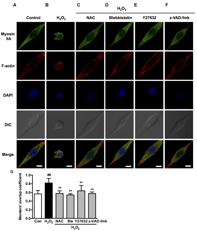
Figure 9.
Caspase, ROCK, myosin II inhibition attenuates H2O2-induced myosin IIA-actin interaction in PC12 cells. (A–F) PC12 cells were untreated or pretreated with 1 μM blebbistatin, 10 μM Y27632 or 10 μM z-VAD-fmk for 1 h prior to 100 μM H2O2 treatment for 12 h. Positive control was treated with NAC (500 μM) for 1 h prior to H2O2 exposure. Myosin IIA (green), filamentous actin (red) and DAPI (blue) were detected by confocal microscope as indicated in Figure 2. Bar, 5 μm. (G) The quantitative co-localization of myosin IIA with F-actin was evaluated on basis of Manders’ overlap coefficients. Results were expressed as mean ± SD (##P < 0.01 vs. control, **P < 0.01 vs. H2O2-treated cells).

