Figure 3.
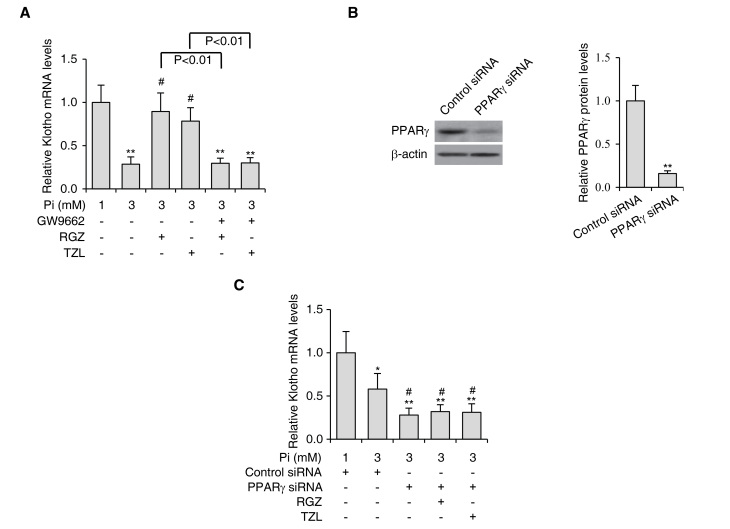
PPARγ agonists enhance Klotho expression in VSMCs. (A) VSMCs were subjected to Pi-induced calcification and treated with RGZ or TZL (10 µM each) in the absence or presence of PPARγ inhibitor GW9662 (10 µM). The expression levels of Klotho were determined by reverse-transcription quantitative polymerase chain reaction analysis. (B) The protein levels of PPARγ in VSMCs treated with siRNA targeting PPARγ were determined by western blot analysis. Quantified PPARγ protein levels are shown in the right panel. β-actin was used as the loading control. **P<0.01 vs. control siRNA-transfected cells. (C) Expression levels of Klotho in VSMCs subjected to Pi-induced calcification treated with RGZ or TZL (10 µM) in the absence or presence of PPARγ siRNA. Values are expressed as the mean ± standard error of the mean of three independent experiments. *P<0.05, **P<0.01 vs. cells in 1 mM Pi group. #P<0.05 vs. cells in 3 mM Pi only group. VSMC, vascular smooth muscle cell; Pi, inorganic phosphate; PPAR, peroxisome proliferator-activated receptor; TZL, thiazolidinedione; RGZ, rosiglitazone; siRNA, small interfering RNA.

