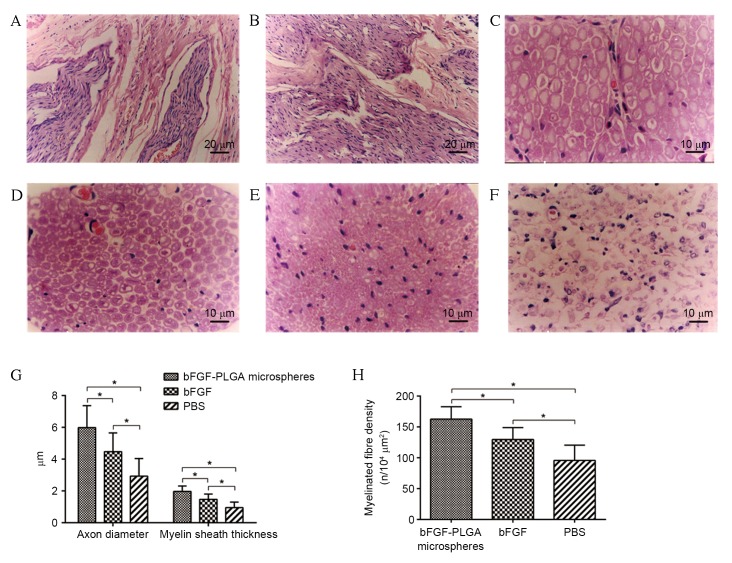
Figure 5.
Histological analysis of the regenerative nerve fibres. (A) At six weeks following surgery, regenerative nerve fibres were growing towards the central area of silicone tube. (B) At 6 weeks post-surgery, the proximal nerve had merged with the distal end in some samples, particularly in the bFGF-PLGA microspheres group. (C) Normal sciatic nerve. At 12 weeks post-surgery the regenerative nerve fibres in the (D) bFGF-PLGA microspheres group were markedly the most dense and best arrayed of the groups, followed by the (E) bFGF and (F) PBS groups, which displayed the lowest degree of axonal regeneration and littered array. (G) Comparisons of the thickness of myelin sheath, axon diameter and (H) density of myelinated fibres, indicating that the bFGF-PLGA microspheres group had the thickest myelin sheath, the largest axon diameter and the highest density of myelinated fibres of the regenerated nerve *P<0.05. bFGF, basic fibroblast growth factor; PLGA, poly-lactic-co-glycolic acid.

