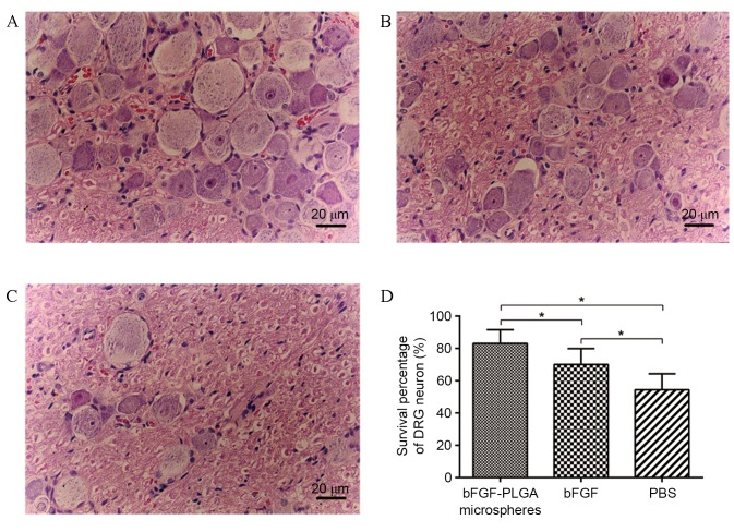
Figure 6.
Neurons in the DRG. (A) In the bFGF-PLGA microspheres group, neurons in DRG were distributed evenly and were almost normal in shape and number. (B) In the bFGF group, the number of neurons in DRG was fewer than those in the bFGF-PLGA microspheres group, although the shape was almost normal. (C) In the PBS group, neurons in DRG were markedly thinner and more sparsely distributed, with irregular cell shape, smaller nuclei and more cluttered nucleoli. (D). The highest survival percentage of DRG neurons was observed in the bFGF-PLGA microspheres group, followed by the bFGF and PBS groups. *P<0.05. DRG, dorsal root ganglion; bFGF, basic fibroblast growth factor; PLGA, poly-lactic-co-glycolic acid.

