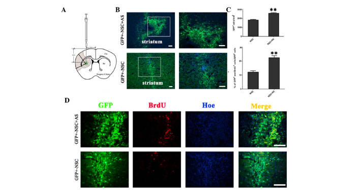
Figure 1.
Astrocytes promote the survival and proliferation of transplanted NSCs in MCAO rats. (A) Schematic drawing of a frontal section through the striatum, illustrating the distribution and dispersion of GFP+ grafted cells. The shaded area indicates the region of ischemia. (B) On day 7 following transplantation, GFP+ NSCs were observed in rats from the co-transplantation group and NSC alone group. (C) A significantly increased number of living cells were observed in rats co-transplanted with AS compared with rats without AS; **P<0.01. (D) BrdU incorporation assays showed that GFP+BrdU+ cells were increased in the co-transplantation group compared with the group without astrocytes. Scale bars, 100 µm. GFP, green fluorescent protein; NSC, neural stem cell; AS, astrocytes; Ctx, cortex; Str, striatum; BrdU, bromodeoxyuridine; Hoe, Hoechst 33342.

