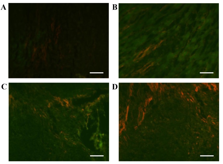
Figure 3.
Differentiation of rat MSCs in infarcted hearts. Cardiac actin immunofluorescence staining of DAPI-labeled MSCs four weeks following transplantation revealed DAPI-positive and actin-positive cells in the infarct area. (A) Infarcted group, (B) chitosan alone, (C) MSCs alone and (D) combined chitosan and MSC treatment. Red represents actin; bars represent 100 µm. MSC, mesenchymal stem cells.

