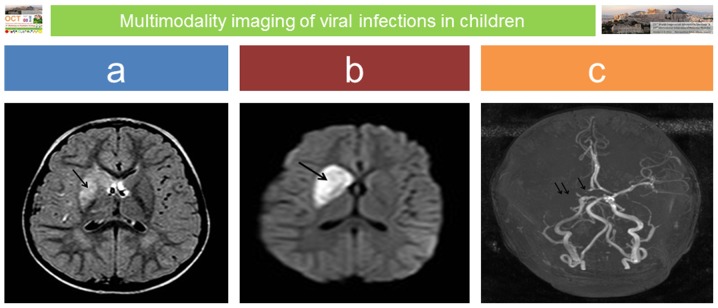
Figure 3.
Post-varicella vasculitis and stroke: a 3-year-old boy presented with acute onset of left upper arm weakness and facial nerve palsy; there was a recent previous history of ‘gastroenteritis’. MRI performed on emergency admission revealed an extensive area of abnormal signal intensity at the region of the right basal ganglia on axial fluid attenuation inversion recovery (FLAIR) image (arrow in panel a), which presented restricted diffusion on diffusion weighted imaging (DWI) sequence (arrow in panel b) in keeping with acute ischaemic infarct. Absence of flow void on magnetic resonance angiography (MRA) was confirmed in the proximal part of the right anterior cerebral artery (ACA) (single arrow in c) and along the right middle cerebral artery (MCA) (double arrow in panel c).

