
Fig. 4.
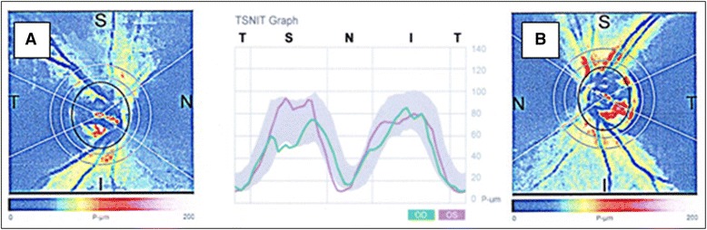
Scanning Laser Polarimetry (GDx ECC). Diminished retinal nerve fibre layer (RNFL) thickness in the superior/temporal peripapillary area, typical for glaucomatous damage in the right eye (a), corresponding with the area of choroidal infarction and the visual field defect. No detectable RNFL damage is present in the left eye (b)