
Figure 2.
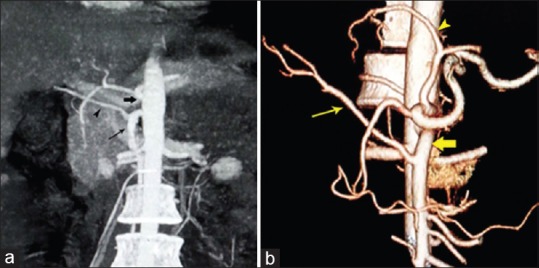
(a) Coronal maximum intensity projection image showing replaced right hepatic artery (arrowhead) from a superior mesenteric artery (thin arrow). Thick arrow points to celiac trunk. (b) Volume-rendered image showing the right hepatic artery (thin arrow) is originated from superior mesenteric artery (fat arrow), also notice to the left hepatic artery off left gastric artery (arrowhead)