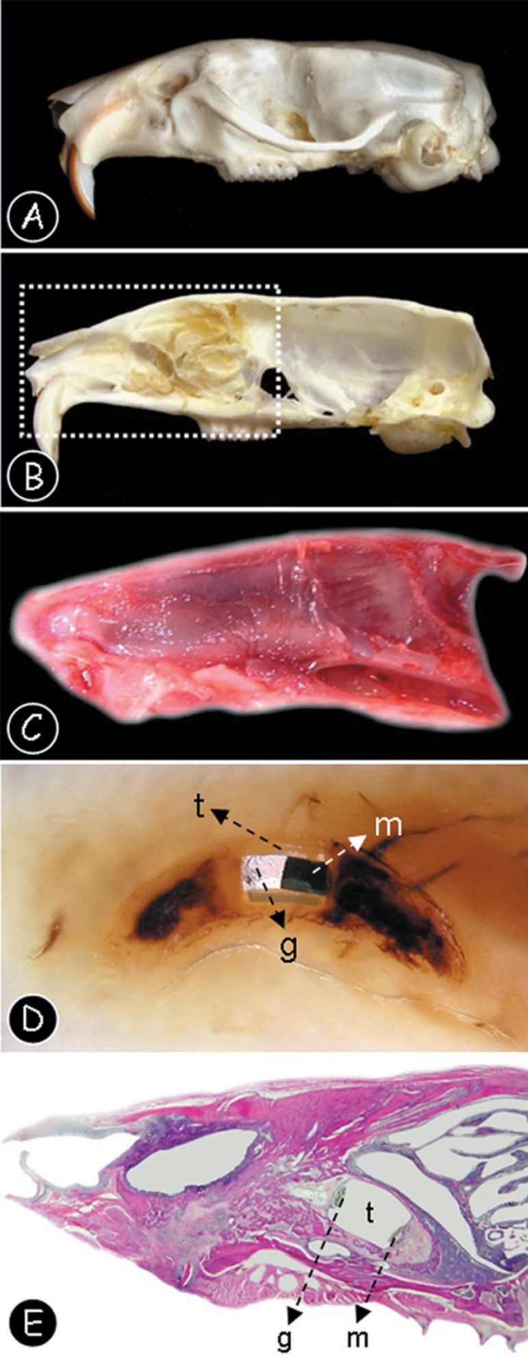
Figure 2.
(A) Lateral view of the rat skull anatomy after maceration procedures. (B) Medium sagital section of the rat skull, after the nasal septum excision (C) Lateral view of the right hemi-maxilla after surgical removal. (D) Longitudinal section of the dental alveolus after paraffin inclusion. Note that the polyethylene tube is present (t), filled with MTA (m) and the sealing material (g). (E) Panoramic tissue section of the right hemi-maxilla after hematoxylin and eosin staining. Note that the polyethylene tube is present (t), filled with MTA (m) and the sealing material (g)

