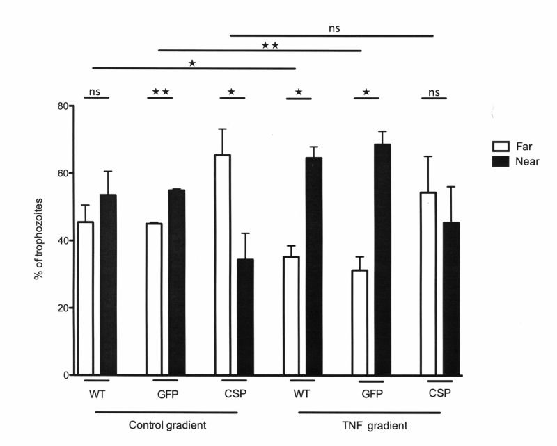
Figure 5. FIGURE 5: In vitro TNF chemotaxis is abolished in CSP-AS trophozoites.

The displacement of the E. histolytica trophozoites was analyzed after 2 h of incubation in the presence of various compounds. Mean numbers of trophozoites in each group (either distal or proximal to the source) were compared in a Student's t-test. In the absence of chemoattractant (incomplete TY medium), WT trophozoites were distributed homogeneously along the coverslip. When the agarose slice was filled with TNF (50 nM), there were significant displacements of WT and GFP trophozoites up the gradient toward the TNF source. In contrast, the presence or absence of a TNF gradient did not significantly affect the distribution of CSP-AS trophozoites. Data are representative of three to four independent experiments. ** P < 0.01; * P < 0.05; ns, not significant.
