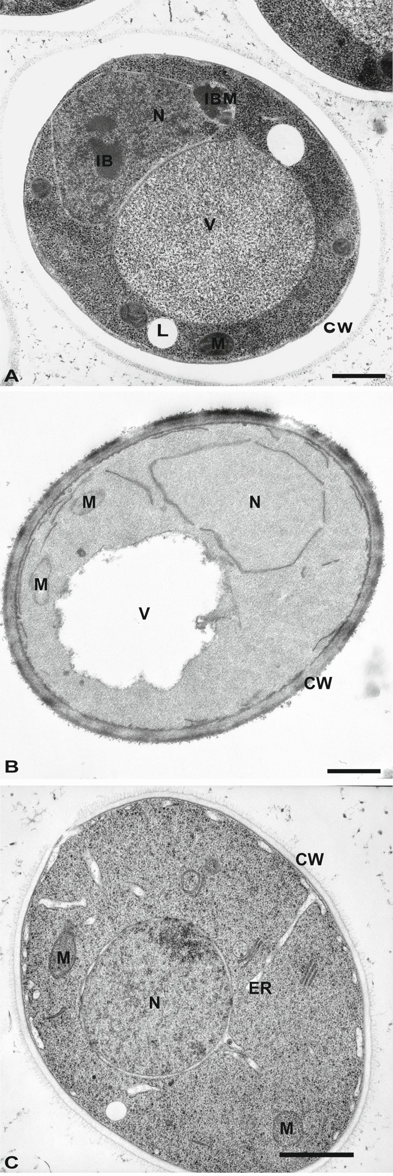
Figure 2. FIGURE 2: Morphology of yeast cells embedded in epoxy resins.

(A) Cells were cryofixed in liquid propane, freeze-substituted in acetone containing 4% OsO4 and embedded in Epon. CW, cell wall; N, Nucleus; IB, Inclusion body; IBM, Inclusion body with membrane; L, lipid droplets; V, Vacuole. Scale bar, 0.5 µm. This image was originally published in 157 © Springer.
(B) Yeast was fixed with 1.5% KMnO4, dehydrated with acetone and embedded in Spurr’s resin. CW, cell wall; M, mitochondria; N, Nucleus; V, vacuole. Scale bar, 0.5 µm. This image was originally published in 158 © the American Society for Biochemistry and Molecular Biology.
(C) Cells were high-pressure frozen, freeze-substituted in acetone, and embedded in a mixture of Epon-Spurr’s resin. CW, cell wall; ER, endoplasmic reticulum; M, mitochondria; N, nucleus. Scale Bar, 1.0 µm. This image was originally published in 26 © Elsevier Limited.
