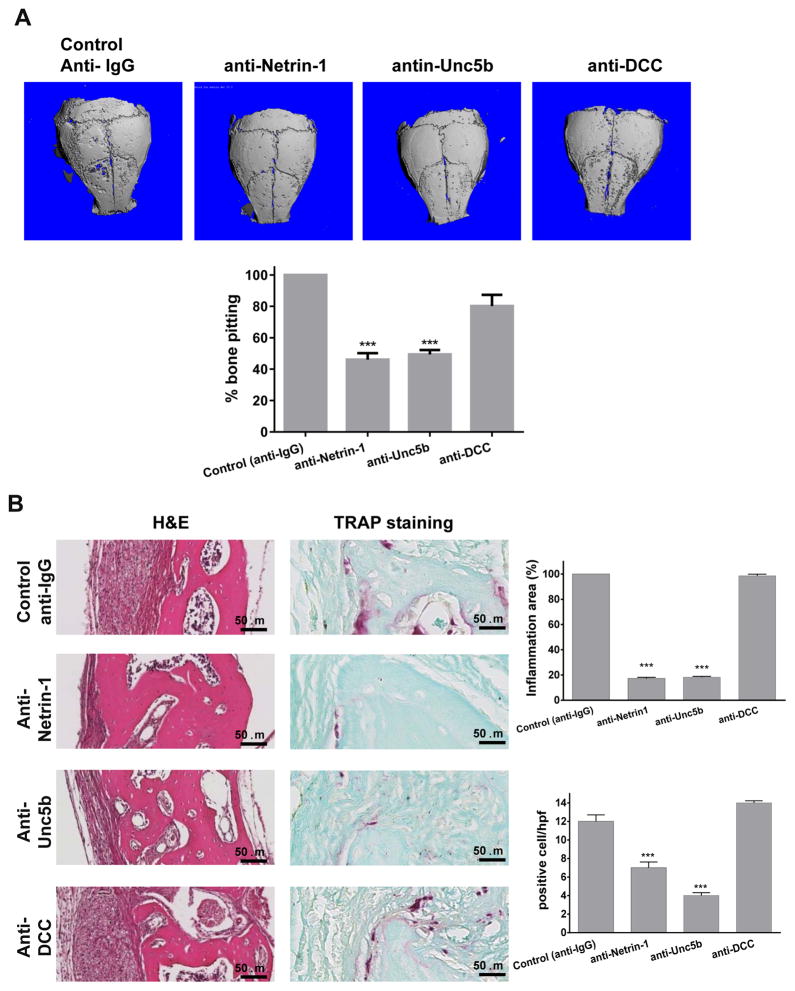
Figure 2. Blocking Netrin-1 using monoclonal antibody injections decreases bone pitting, inflammation and osteoclasts in mouse calvariae after exposure to wear particles.
A) The figures show representative microCT images of calvaria of mice treated with UHMWPE wear particles combined with anti-IgG (Control), anti-Netrin-1, anti-Unc5b or anti-DCC blocking antibodies at 10μg (n=5 mice per group). B) Calvariae were stained with hematoxylin & eosin to determine the presence of inflammation on the outer bone surface. The area of inflammatory infiltrate was quantified and expressed as a percentage of the area of the control particle-exposed mice (n = 5 mice per group). Shown are representative images for TRAP staining for osteoclasts in mice calvariae and the mean (±SEM, n=5 mice per group) number of osteoclasts/hpf. Images were taken at 400X magnification. Scale bar indicates 50 μm. Data were expressed as mean±SEM (n=5 per group). * p<0.05, **p<0.001, ***p<0.001 compared to control (ANOVA).

