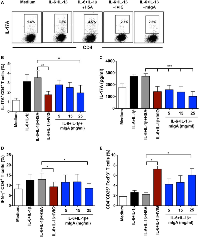
Figure 2.
Monomeric IgA (mIgA) reciprocally regulates Th17 cells and FoxP3+ T cells under Th17 amplification conditions. (A) Flow cytometry analysis of intracellular IL-17A in the memory CD4+ T cells cultured in serum-free X-vivo medium in the presence of anti-CD3 and anti-CD28 mAbs alone (medium) or stimulated with IL-6 and IL-1β for 6 days. mIgA (25 mg/ml), IVIG (25 mg/ml), or human serum albumin (HSA) (10 mg/ml) (0.15mM) were added to the T cell cultures after 12 h of cytokine stimulation. Data from one of five independent experiments are presented. (B) Percentage of IL-17A+CD4+ T cells (mean ± SEM, n = 5 donors) and (C) amount of secreted IL-17A (mean ± SEM, n = 9 donors) in T cells cultured under above conditions. mIgA was added at three different concentrations (5, 15, and 25 mg/ml). (D) Percentage of IFN-γ+CD4+ T cells (mean ± SEM, n = 5 donors) and (E) CD4+CD25+FoxP3+ T cells (mean ± SEM, n = 5 donors) among CD4+ T cells cultured under above conditions. Statistical significance as determined by one-way ANOVA is indicated (*P < 0.05, **P < 0.01, ***P < 0.001).

