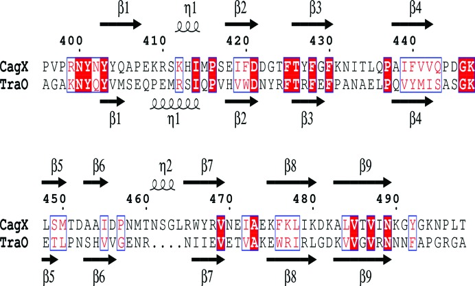
Figure 5.
Anmino-acid sequence alignment of CagXct and TraOct. The secondary-structure elements are indicated above and below the alignment, respectively, as β-strands and η-helices (310-helices). Conserved residues are shown in red boxes; matching amino acids with similar properties are shown in blue boxes. The secondary-structure assignments were carried out and the figure was produced using ESPript3 (Robert & Gouet, 2014 ▸).

