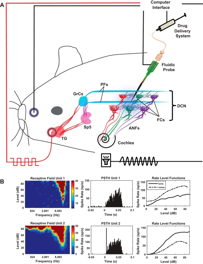
Fig. 1.
Schematic of the experimental setup. A: auditory stimulation delivered to the guinea pig cochlea through a hollow ear bar activates the auditory nerve fibers (ANFs) and DCN fusiform cells via ANF synapses on fusiform cell basal dendrites. Electrical pulses on the face activate the somatosensory pathway to the DCN by stimulating the trigeminal ganglion (TG) and spinal trigeminal nucleus (SP5) projections to the granule cells (GrCs), which relay their output to the fusiform cells via parallel fibers (pfs) that synapse on their apical dendrites. For bimodal stimulation, somatosensory stimulation is delivered in precise temporal proximity with the auditory stimulation. The neural activity is recorded with a fluidic probe. Baseline fusiform cell activity and plasticity were evaluated before and after delivery of a mAChR blocker (atropine, 80 µM, 2 µl, 100 nl/min) in the fusiform cell layer. B: examples of characteristic responses of fusiform cells: receptive fields, peristimulus time histograms, and noise and tone rate-level functions were recorded before the experiment to characterize the type of the fusiform cells. Shown are 2 examples of the most common unit types recorded, a build-up type III (top row) and a pause build-up type I (bottom row).

