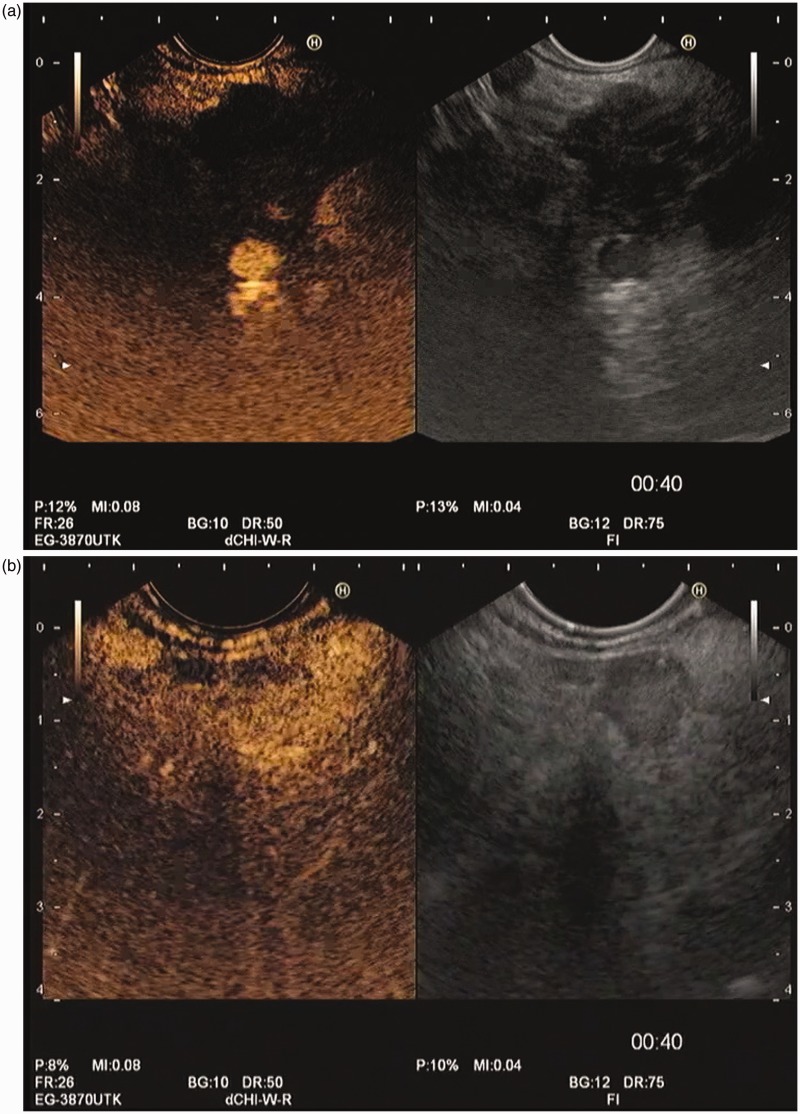
Figure 4.
Contrast-enhanced harmonic endoscopic ultrasound evaluation of pancreatic solid masses, both at the end of arterial phase, beginning of venous phase (00:40 minutes). (a) Pancreatic adenocarcinoma, showing the typical hypovascular pattern. (b) Neuroendocrine pancreatic tumor, showing a hypervascular pattern.

