Fig. 8.
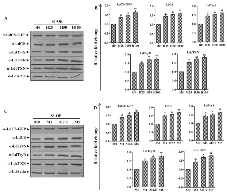
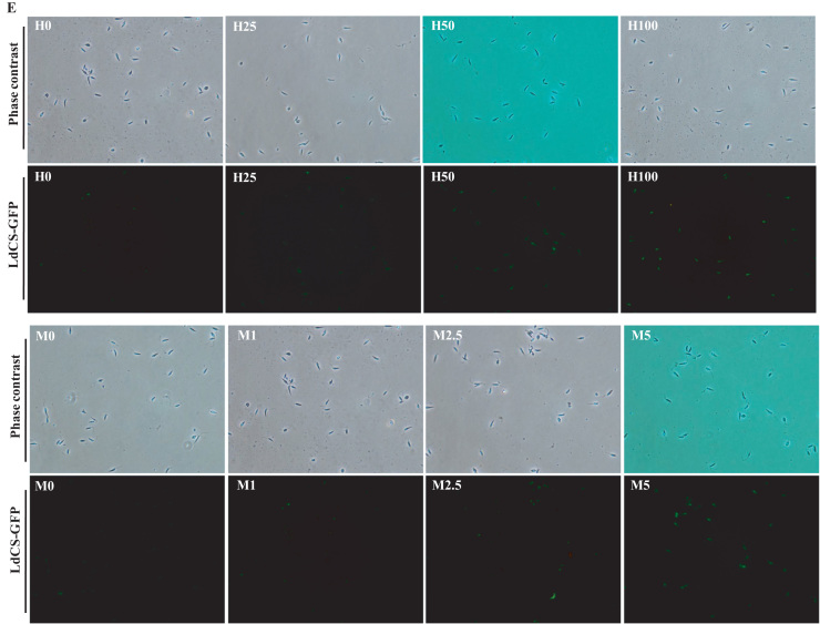
LdCS over-expression primed modulation of endogenous LdCS as well as thiol pathway proteins (LdTryS, LdTryR and LdcTXN) in L. donovani under oxidative stress. (A and C) Total cell lysates prepared from S1-OE parasites exposed to lethal doses of H2O2 (25 µM, 50 µM and 100 µM) and menadione (1 µM, 2.5 µM and 5 µM) for 6 h were separated on 10% SDS-PAGE and subjected to western blot analysis using α-LdCS, α-LdTryS, α-LdTryR, α-LdcTXN and α-LdActin antibodies, respectively. (B and D) Relative band intensities were determined by densitometric analysis and fold change in the expression of LdCS-GFP, LdCS, LdTryS, LdTryR and LdcTXN in S1-OE parasites exposed to lethal doses of H2O2 and menadione were graphically represented as means±SEM of three independent sets of experiments. Untreated parasites were used as a control in the experiment. *, p<0.05; **, p<0.01; by Student's t-test using GraphPad Prism 5.0. (E) Fluorescence microscopy image of S1-OE parasites over-expressing LdCS-GFP protein treated with H2O2 (25 µM, 50 µM and 100 µM) and menadione (1 µM, 2.5 µM and 5 µM) for 6 h. Untreated parasites were used as a control. ,.


